
7 Steps to MAXIMIZE Skin Biopsy Results
Dr. William Fortney
Dermatological conditions are among the most frequent presentations to companion animal practices. They are also often frustrating problems to manage. Identifying a definitive diagnosis can help point the way to a cure or better maximize a long-term management strategy.
Following a good history, a thorough physical examination, and multiple skin scrapings, a skin biopsy is often the next logical diagnostic step. At best, a quality skin biopsy can establish a definitive diagnosis of the condition… at the least, a biopsy results will provide a likely list of differential diagnoses. However a skin biopsy takes time, money and some knowledge to maximize the results. A skin biopsy is a valuable diagnostic tool. However, like all the dermatology tools; it may not always provide the definitive diagnosis.
The following 7 Steps will help you MAXIMIZE a Skin Biopsy Results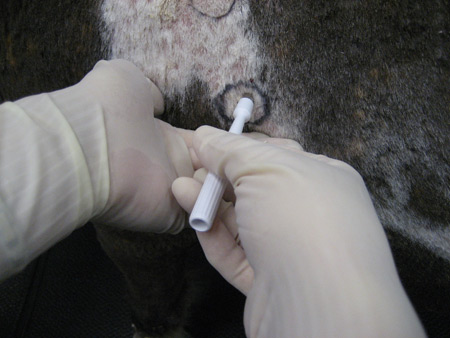
1. Always biopsy the best lesion(s) and the best part of each lesion:
An early lesion is better than old one; an untreated lesion is better than treated one; biopsies from several different lesions are better that one mega lesion; and excoriated lesions (scratched, licked, or chewed) are usually not very helpful.
2. Take multiple biopsies when possible:
Even on a single lesion, multiple small samples are better than one big one. Depending on the disease, the definitive diagnostic finding is not always present in a single biopsy; therefore, multiple biopsies greatly increase the probability of obtaining an accurate diagnosis.
3. Do Not Prep the biopsy area:
Unlike most procedures, preparation of the biopsy site may destroy important pathologic findings by removing crust, scale, exudate, pustules, vesicles, mites, yeast, and even superficial bacteria.
4. Use the proper instruments:
A sharp 8 mm diameter skin punch biopsy or scalpel blade is the best option. When collecting the biopsy, NEVER use laser or electro-cautery as these instruments burn / damage the delicate sample tissue.
5. COMPLETELY fill out the submission form:
The more information contained in the form the better. The signalment, history, lesion description and location, and additional testing all help the pathologist better interpret the histopathology findings.
6. Digitally documenting the lesion:
When possible, please send a color print, CD, or email the picture to receiving@vet.k-state.edu . "A picture is worth a 1000 words" compared to the written the description on your submission form. A pic is even more important if you are using the KSVDL Dermatology Service were the dermatologist is often present. Note: make sure the picture is labeled with the practice name and the patient is identified.
7. Use the KSVDL "Dermatology Service":
Once a week, the KSVDL pathologists and the KSU Veterinary Health Center dermatologist review most of the previous week’s skin biopsy cases submissions. By requesting "KSVDL Dermatology Service", most of the pathologists and a Board Certified dermatologist will review the submission form, any forwarded pictures, and the histopathology slides. The Dermatology Group develops a consensus on the probable diagnosis(s), additional recommended diagnostics and/or various management options. While the advantages of this service are obvious, the once weekly Derm Group meeting does slow the turnaround time. For more information on skin biopsies, go to Test and Fees or contact KSVDL Client Care at 866-512-5650 or clientcare@vet.k-state.edu.
Next Article : General Sample Shipping Preparation
Return to Index