
January 2019
Canine Ischemic Dermatopathy: Part I
Canine familial dermatomyositis
 |
| Table 1 |
By Drs. Charan Ganta and Sarah Schneider
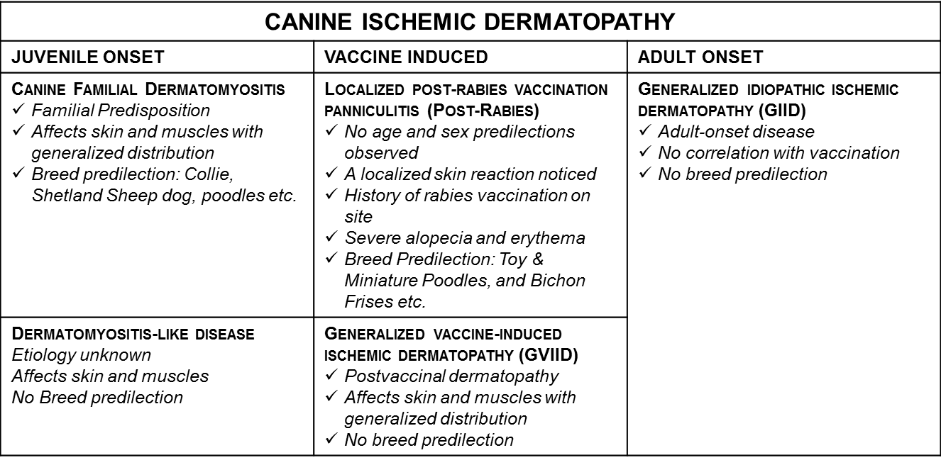
Canine ischemic dermatopathies include a group of vasculopathic skin diseases that result in decreased oxygenated blood supply to the skin. These include juvenile and adult onset types (Table 1).
Canine familial dermatomyositis is a juvenile onset ischemic dermatopathy and myopathy that is most commonly reported in Collies, Shetland Sheepdogs, Beaucerons, Belgian Tervurens and Portuguese Water Dogs.
Clinical Manifestation
The clinical signs manifest typically between 1.5 to 6 months of age. The disease initially shows signs of alopecia, erythema, crusting, erosions, ulcerations, progressing to scarring alopecia and hyper or hypopigmentation with sloughing of skin, usually on the bony prominences. Often these lesions are complicated by secondary infections due to immune suppression. Dermatitis is not often associated with myositis, but when present, the masticatory muscles are most commonly affected. Peripheral muscle involvement can lead to generalized weakness. Megaesophagus is another complication, but is rarely reported.
|
|
|
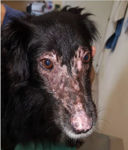
Figure 1. Collie mix female dog. Bilateral, non-symmetric alopecia with scarring and hypopigmentation on the dorsal skin of the nose with involvement and multifocal erosions of the nasal planum. Photo courtesy of ISVD. |
Etiology and Pathogenesis
Due to a strong breed predilection, a genetic component to this disease is believed to be present. The pathogenesis of dermatomyositis remains unknown; the presence of antigen-antibody complexes in the vessel walls suggests a type III hypersensitivity reaction. In addition, elevated serum complement levels and class G immunoglobulins have also been demonstrated.
Disease Diagnosis and Sample Collection
The diagnosis of dermatomyositis is based on typical clinical signs, age of onset, breed predilection and distribution with further confirmation by histopathology. Often the disease can be complicated by secondary conditions like pyoderma, demodicosis, dermatophytosis and Malassezia and therefore multiple deep skin biopsy sections from different locations should be submitted along with a complete clinical history. Often the disease diagnosis is complicated by co-existing allergic dermatitis.
Histopathological Features
The histologic lesions, which are common in all conditions of canine ischemic dermatopathy, are characterized by cell-poor vasculitis (cause vascular compromise and cutaneous ischemia). The vascular changes include loss of endothelial cells, with hyalinization and occasional leukocytoclasia. Altered staining of the collagen, fading atrophy of the follicles is most frequently noticed. In advanced cases, dermal-epidermal Vacuolation, pigmentary incontinence and artefactual lifting of the epidermis could occur. Muscular lesions when noticed include mixed inflammation, regeneration, fibrosis, and atrophy.
Prognosis
Canine familial dermatomyositis is progressive and management is often challenging and dependent on the stage and severity of the disease and the presence of secondary infections. For case management advice please call the Veterinary Health Center at the Kansas State University College of Veterinary Medicine for a consultation with the clinical dermatologist.
For any additional questions and references related to this newsletter please contact KSVDL Client Care at 866-512-5650 or clientcare@k-state.edu.
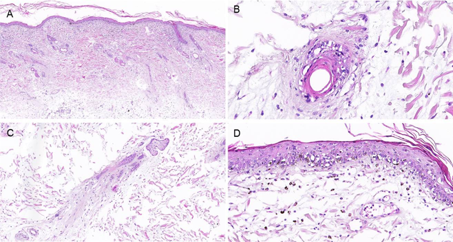 |
Figure 2. H&E sections. A. Diffuse and moderate to severe follicular atrophy with homogenization and pallor of dermal collagen. B. Cell poor vasculitis with vascular endothelial hyalinization. C. Marked follicular atrophy and pale staining dermal collagen. D. Dermal-epidermal junction showing cell-poor lymphocytic infiltration, with basal cell Vacuolation and pigmentary incontinence. Photo courtesy of Dr. Charan Ganta (KSVDL) and Dr. Diane Longfellow (Grassland Veterinary Hospital |
NEXT: Bovine Abortion Workups