
The Diagnosis of Fungal Kerion in Dogs
Dr. Gordon Andrews and Dr. Fortney
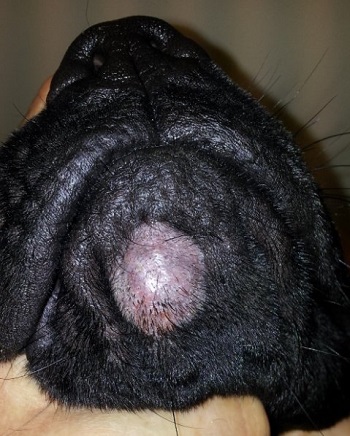
Figure 1: Raised hairless cutaneous nodule on the chin of a dog diagnosed as a fungal kerion. |
Although often clinically over-diagnosed, the typical dermatophyte infection is still a fairly common skin disease seen in dogs. A less common nodular form of dermatophyte infection is a fungal kerion. Because the fungal kerion has a non-typical dermatophyte infection appearance, the precise diagnosis is often elusive and easily missed.
Infection is usually caused by M. gyseum or T. mentagrophytes. The dermatophytes are located deep within the dermis and may be few in number, so routine diagnostic tests such as a Wood’s lamp examination, microscopic examination of hair shafts for fungal elements, and fungal culture often yield negative results. The presence of secondary bacterial infection (Staph. sp) may complicate the diagnostic findings.
Grossly a fungal kerion is a firm to boggy, well–circumscribed, raised, focal or multifocal cutaneous nodule (Figure 1). Occasionally the lesion is exudative and may have draining tracts. Fungal kerions can occur anywhere on the body, but most commonly are localized on the face, pinnae, paws and/or tail. Depending on the lesion appearance, location, and number of kerion(s) involved; a fungal kerion can mimic bacterial furunculosis, demodex, histocytoma or other cutaneous neoplasia, or even auto-immune disease.
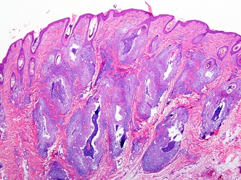
Figure 2: Multifocal areas of pyogranulomatous inflammation oriented around and replacing hair follicles. |
Biopsy DX
Because these lesions typically present as a single cutaneous mass, neoplasia is suspected and they are frequently surgically excised and submitted for histopathologic examination. Histologically the lesion is characterized as a nest of ruptured hair follicles replaced by suppurative to pyogranulomatous inflammation sometimes with eosinophils oriented around hair fragments that contain fungal hyphae and are surrounded by fungal spores (Figures 2 and 3).
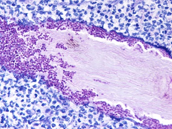
Figure 3: Hair shaft with dermatophyte hyphae within the hair shaft and fungal spores surrounding the hair. The hair shaft is surrounded by neutrophils and macrophages. |
With a single and uncomplicated kerion, the use of a topical “antifungal” agent may be sufficient therapy. However complicated and multiple lesions are best managed with both topical and systemic “imidazole” medication. The secondary bacterial infections should also be managed. Even with appropriate treatment strategies, it may take four – eight weeks for the lesion to resolve. Rarely the infected hair follicles are sufficiently damaged and never re-grow.
Next Article: Canine and Feline Core Vaccine Titer Screening
Return to Index