
March 2020
Confirmed Cases of Cache Valley Virus and Related Viruses at KSVDL
By Dr. Giselle Cino
BACKGROUND
We have now confirmed two cases of Cache valley virus (CVV) infection in small ruminants, as well as other suspected cases at KSVDL. CVV and other related viruses are mosquito-borne bunyaviruses of the serogroup Bunyamwera, which are endemic in North America and can cause reproductive disease in sheep and goats.
CVV is considered the most economically important virus of the serogroup and the most common cause of large reproductive losses in the sheep industry in North America. Cases in the state of Kansas are sporadically reported, but epizootics can occur in naïve herds. Currently, there are no available vaccines or treatment for the disease. The disease is zoonotic and transmitted to humans only by mosquito bites and not by direct contact with infected animals.
|
|
PATHOGENESIS
The virus infects kids and lambs in utero and leads to pregnancy failure and abortions of offspring with congenital malformations, if the infection occurs between 28- 48 days of gestation. The disease is transmitted by mosquitoes of multiple families including Aedes, and culicoid midges. The virus does not cause any clinical signs in the dam, leading to difficulty in identifying carriers of the virus in the herd. Following a short viremic phase, the dam mounts an antibody response to the virus and clears the virus. “Open” dams due to embryonal loss and abortion, due to malformations in musculoskeletal and central nervous system (CNS) systems, are the major clinical manifestations of the disease.
LESIONS AND DIAGNOSIS
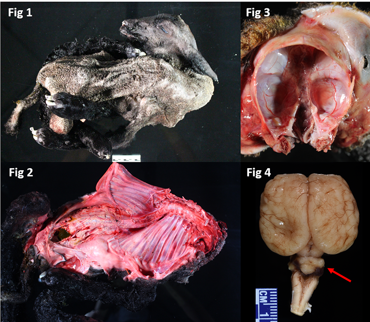
On necropsy, the fetuses show arthrogryposis (Figure 1) characterized by curled, twisted, and crooked limbs. There could also be deviation of the spinal cord (scoliosis) or torticollis (Figure 2). CNS changes include varying degrees of hydranencephaly (Figure 3) and cerebellar aplasia or hypoplasia (Figure 4, red arrow), and generalized muscle atrophy. Testing of fetal tissues (including placenta and brain) can lead to isolation of the virus and diagnosis of the disease. Serologic analysis of dams aids in detecting the seroprevalence of the virus in the herd, but is not confirmatory of the disease in fetus.
DIFFERENTIAL DIAGNOSES
Although CVV is a cause of major congenital malformations and abortions in small ruminants, several other conditions can have similar clinical presentation. Among these differentials are the ingestion of teratogenic plants during gestation, such as Lupinus spp., and in utero infection with bluetongue virus, bovine viral diarrhea virus and border disease virus. Finally, some skeletal deformities caused by inherited genetic defects, such as Spider Lamb Syndrome (chondrodysplasia), can also appear grossly similar to arthrogryposis.
Therefore, testing the herd for detection of these infectious pathogens and teratogenic plants is imperative for a correct diagnosis and management of the disease, as well as for the establishment of adequate preventative measurements.
TAKE HOME MESSAGES
Cache valley virus is a mosquito-borne disease that infects lamb and goat in utero, leading to pregnancy failure, abortions and congenital malformations in fetuses. Infected dams show no clinical signs or lesions, making it challenging to identify carriers in the herd. There is no vaccine or treatment available for the disease. Serology testing on dams and full necropsy and sampling of the aborted fetuses (including brain and placenta) are needed to determine the disease status in the herd, as similar congenital malformations can be observed with other infectious, genetic and ingestion of teratogenic plants.
References:
- Aline Rodrigues Hoffmann, Piotr Dorniak, Justyna Filant, Kathrin A. Dunlap, Fuller W. Bazer, Andres de la Concha-Bermejillo, Christabel Jane Welsh, Patricia Varner, John Francis Edwards. Ovine Fetal Immune Response to Cache Valley Virus Infection. Journal of Virology Apr 2013, 87 (10) 5586-5592.
- Lisa Waddell, Nicole Pachal, Mariola Mascarenhas, Judy Greig, Shannon Harding, Ian Young, Barbara Wilhelm. Cache Valley virus: A scoping review of the global evidence. Zoonoses Public Health. 2019;66:739–758.
- Victoria B. Ayers, Yan‑Jang S. Huang, Amy C. Lyons, So Lee Park, James I. Dunlop, Isik Unlu, Alain Kohl, Stephen Higgs, Bradley J. Blitvich, Dana L. Vanlandingham. Infection and transmission of Cache Valley virus by Aedes albopictus and Aedes aegypti mosquitoes. Parasites Vectors (2019) 12:384
Giselle Cino, DVM, PhD, ACVP is an anatomic pathologist in the Kansas State Veterinary Diagnostic Laboratory
NEXT: Cytologic Evaluation of Peripheral Lymph Node Aspirates
Return to Index