
November 2017
An Injection Site Reaction: Post-Rabies Vaccination Panniculitis in a Toy Poodle
By Dr. Charan Ganta
|
||
|
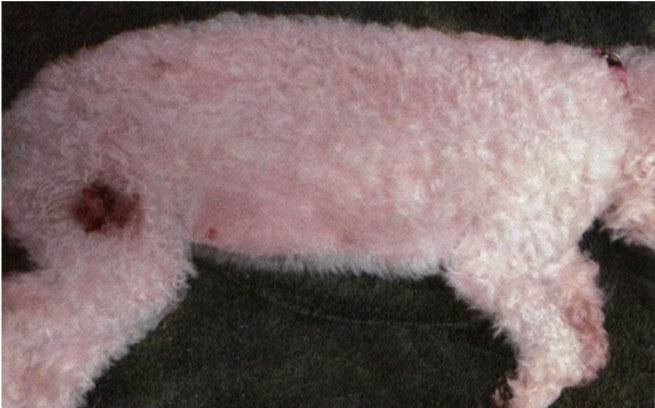
Figure 1. Post rabies vaccination panniculitis in a Toy Poodle. Alopecia with hyperpigmented area developed at the site of prior subcutaneous rabies vaccination above the right hip area. Image courtesy of Dr. Darla Dwyer, Countryside Vet Clinic. |
||
|
||
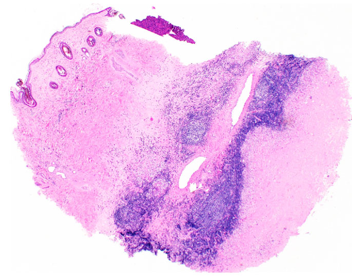
| Figure 2. Post rabies vaccination panniculitis with characteristic nodular aggregates of perivascular lymphocytes in the subcutis with follicular atrophy in the dermis. | ||
|
||
|
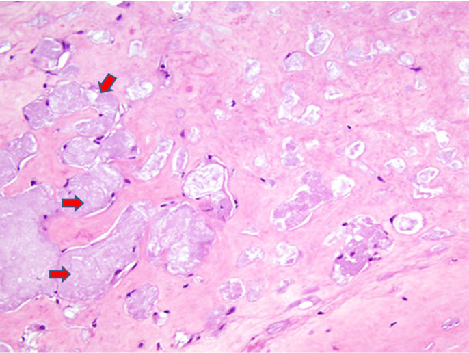
Figure 3. Necrosis surrounds foci of amphophilic homogeneous extracellular, intravascular and intrhistiocytic material (vaccine product). Arrows represent vaccine material. Image source: askjpc.org |
What is Post Rabies Vaccination Panniculitis?
An injection site reaction secondary to post-rabies vaccination is a relatively common canine skin disease often characterized by a focal area of hair loss, thickened skin with minimal gross inflammation. In one study it was reported that the presence of rabies viral antigen in the walls of cutaneous vessels could cause vasculitis and ischemic dermatopathy (2).
Breeds of Dogs Affected
This condition is most frequently diagnosed in Toy or Miniature Poodles and Bichon Frises, as well as other long-haired small breeds are affected with less frequency which include Shih Tzu, Lhasa Apso, Maltese, Silky Terrier, Yorkshire Terrier, Chihuahua, Toy Manchester Terrier, American Eskimo, Poodle crossbreeds, and Miniature Dachshunds. The susceptibility of these breeds had been attributed to enhanced genetic susceptibility and a long anagen hair growth cycle. This condition was very rarely reported in large breed dogs with only one case reported to date.
Clinical Presentation
Clinically, the time interval between vaccination and observation of the lesion was generally 2 to 4 months, but the interval may be longer. The lesion manifests slowly as a variably-sized area of alopecia with irregular margins that subsequently become erythematous, scaly, and hyperpigmented. A small subgroup of dogs may develop widespread alopecia and skin lesions which is called “generalized vaccine-induced ischemic dermatopathy”
Biopsy Sample Collection and Histopathology
A deep biopsy sample of the affected epidermis and subcutis just within the outer margin of the lesion is recommended, often samples collected from the central affected area is of less diagnostic value.
Histopathological examination often reveals dermal pallor, smudging, and follicular atrophy in the superficial dermis and moderate to severe nodular perivascular accumulations of lymphocytes, fewer histiocytes, and occasional plasma cells in the deep dermis and panniculus (figure 2). Occasionally, an amorphous basophilic foreign material presumably constituting vaccine product (figure 3) can be noticed histologically within the deep macrophages. Eosinophils are usually present in reactions due to other types of vaccines, but are inconspicuous in post-rabies vaccination.
Precautions:
Revaccination with subcutaneous rabies vaccine is not recommended, as the syndrome may exacerbate in response to further antigenic exposure. Therefore, for a dog with prior history of post-rabies vaccination panninulitis, it is advisable to use rabies virus vaccines that require administration every three years. It is also advised to avoid administration of multiple vaccines at one time, and inject the vaccines intramuscularly rather than subcutaneously. Spontaneous hair regrowth may occur but can take up to 1 year and may be associated with altered pigmentation. In this case, the skin lesion was surgically removed and the patient recovered completely and doing well.
Reference:
1. Gross TL, Ihrke PJ, Walder EJ, et al. Diseases of the panniculus. In: Skin diseases of the dog and cat. 2nd ed. Ames, Iowa: Blackwell, 2005;538–558.
2. Wilcock BP, Yager JA. Focal cutaneous vasculitis and alopecia at sites of rabies vaccination in dogs. J Am Vet Med Assoc 1986;188:1174–1177.
3. Medleau L, Hnilica KA. Cutaneous vasculitis. In: Small animal dermatology: a color atlas and therapeutic guide. 2nd ed. Philadelphia: WB Saunders Co, 2001;216–217.
Acknowledgements:
Dr. Darla Dwyer and Dr. Amanda Allison; Countryside Vet Clinic
BRD Antimicrobial Resistance Trends are Updated!
Return to Index