
October 2020
FAKE OUT! Hemoparasite or Blood Smear Artifact
By Drs. Brandy Kastl and Nora Springer
Hemoparasites are a differential diagnosis for animals with anemia. Blood film examination is a rapid and cost-effective method to evaluate for hemoparasites. However, numerous artifacts commonly seen on blood smears can mimic hemoparasites. The following quiz is to test your knowledge differentiating hemoparasites from common artifacts and provide tips and tricks for differentiating these entities. (Answers at the bottom of the page!) All images are stained with Modified-Wright stain.
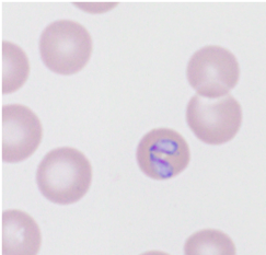
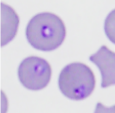
1. Which image below depicts Babesia canis piroplasms inside a canine red blood cell (100x oil objective)?
|
|
|
| A. | B. |
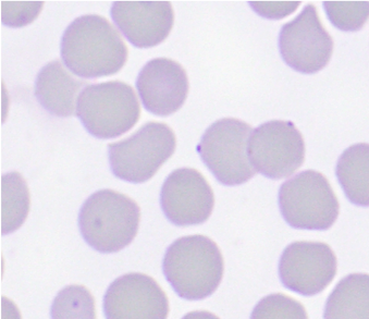
2. Which picture below depicts hemotropic Mycoplasma on feline red blood cells (100x oil objective)?
|
|
|
| A. | B. |
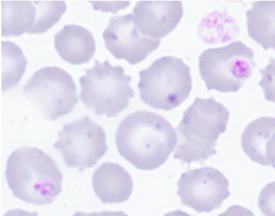
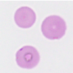
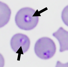
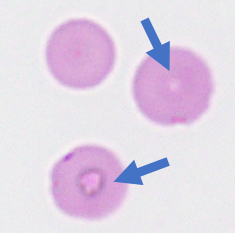
3. Which picture below depicts Cytuaxzoan felis piroplasms in feline red blood cells (100x oil objective)?
|
|
|
| A. | B. |
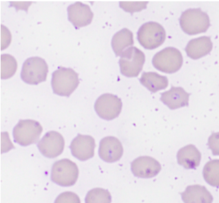
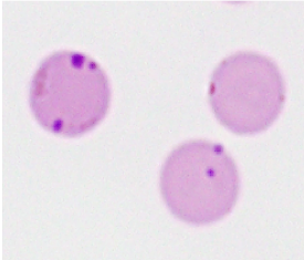
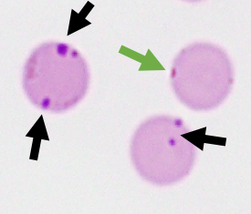
4. Which picture below depicts Anaplasma marginale organisms on bovine red blood cells (100x oil objective)?
|
|
|
| A. | B. |
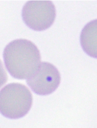
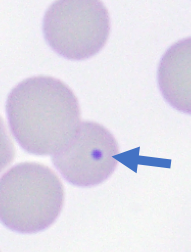
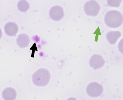
5. Which arrow below depicts Mycoplasma wenyonii organisms in a bovine blood film (100x oil objective)?

|
Answers:
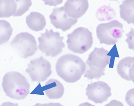
1. B is the image of Babesia canis piroplasms inside a canine erythrocyte. These organisms are round, tear drop, or sometimes ameboid shaped with a single nucleus often to the peripheral margin of the organism (black arrow). Image A shows multiple normal canine platelets (blue arrows) superimposed over canine erythrocytes. Notice the regular distribution of the platelet granules and the variation in platelet size.
|
|
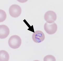
2. B is the image of hemotropic Mycoplasma organisms on the surface of a feline erythrocyte. Notice the consistent size, placement, and staining intensity of the Mycoplasma organisms along the peripheral margin of the erythrocytes (black arrows). Image A shows stain precipitate on the erythrocytes and unevenly distributed throughout the slide background (blue arrows). Notice the irregularity and variation in shape and location on the erythrocytes. Regularly changing rapid stains at least once monthly or more often as needed can reduce this artifact.
|
|
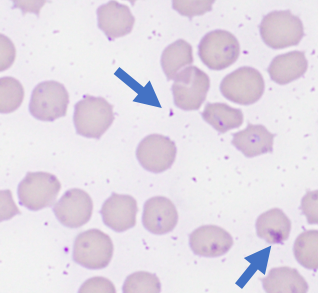
3. A is the image of Cytauxzoon felis piroplasms inside feline erythrocytes. Notice the classic teardrop and rounded shape with a peripheralized dark, crescent-shaped nucleus. Image B shows water artifact on the erythrocytes (blue arrows). This artifact can be of any shape and on any location in the red cell. It can also line the peripheral margin of the cells mimicking either Anaplasma or Mycoplasma spp. When the fine focus is slightly adjusted on this artifact it will change from a dark color to clear, bright, and refractile as the light from the microscope bends through the water droplets. Allowing slides to dry completely prior to staining can eliminate this artifact. Using a hairdryer on the cool or low setting can aid in rapidly drying blood smears and eliminating pesky water artifact.
|
|
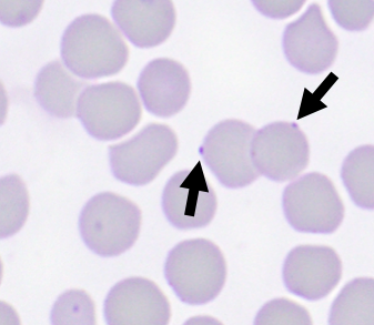
4. A is the image of Anaplasma marginale organisms in bovine erythrocytes (black arrows). Anaplasma marginale appear as prominent, deep purple structures along the erythrocyte margin. Relative to Mycoplasma spp. organisms, Anaplasma marginale is a bit lager and denser. The green arrow in image A denotes water artifact along the margin of the erythrocyte which can sometimes mimic Anaplasma marginale See answer to question 3 for information on water artifacts. Image B is of a Howell-Jolly body, which are nonpathogenic erythrocyte nuclear remnants. These structures tend to stain blue rather than purple, are more regularly round versus Anaplasma marginale, and usually within the erythrocyte rather than along the margin of the cells.
|
|
5. Large numbers of wenyonii(green arrow) are seen free in the background of the smear. Very few Mycoplasma wenyonii organisms adhered to red blood cells is a common finding. The organisms are small (<0.5um) pale lavender to grey and present in cocci, ring and rod-shaped forms. At times, the granules in ruminant platelets (black arrow) can mimic these organisms, particularly when platelets are clumped or pale-staining. Platelet granules are a pink-purple, more regularly spaced, and more variable in size versus Mycoplasma wenyonii organisms.
|
Brandy Kastl, DVM, DACVP is a clinical pathologist in the KSVDL. Nora Springer, DVM, PhD, DACVP is a clinical pathologist and the Section Head for the Clinical Pathology Laboratory and the Immunology Laboratory within the KSVDL.
Next: New Tests at KSVDL
Return to Index