Bovine Summer Pneumonia
By Drs. Giselle Cino, Pankaj Kumar, Kelli Almes and Gregg Hanzlicek
Summer pneumonia, also known as pasture pneumonia is a disease most often observed in preweaned calves on pasture late in the summer. Pathogens that have been most often associated with this disease include bovine coronavirus (BCoV) and bovine respiratory syncytial virus (BRSV). Recently, a Kansas herd experienced the disease early in the spring, shortly after pasture turnout.
History: Approximately 200 commercial beef cows calved in the spring. Calves were administered a respiratory vaccine and a multivalent clostridial toxoid at three days of age. The herd was then housed in a dry lot until the middle of May with no major health issues reported. At that time, calves (approximately 4 months of age) were administered with the same vaccine products received as neonates, and they were moved with their dams to multiple pastures.
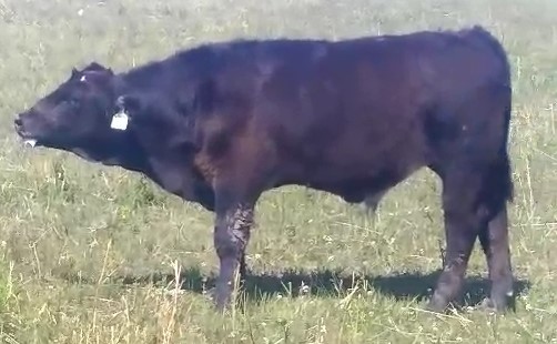
Clinical signs: Approximately three weeks later, calves in multiple pasture locations began to show signs of severe respiratory disease. The clinical signs included ≥106° rectal temperature and extreme dyspnea with open-mouth breathing. (Figure 1, affected calf from this case)
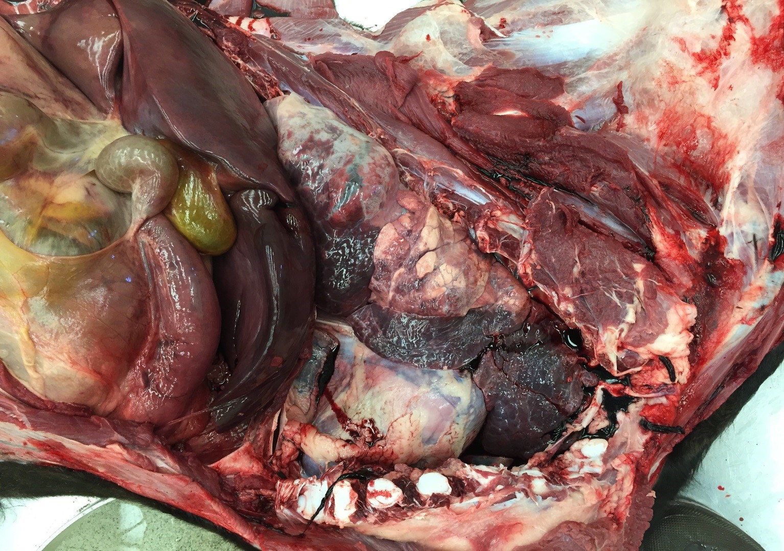
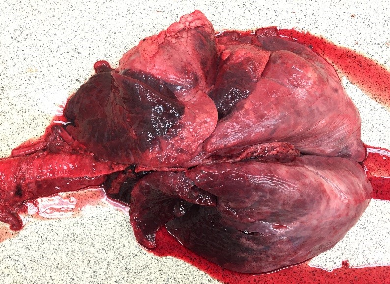
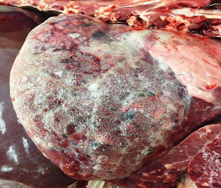
Diagnostics: Three calves were necropsied and histopathology and bovine respiratory panel PCR tests were performed. Gross necropsy description included bronchopneumonia, with severe emphysematous bullae. (Figure 2, 3, 4) Histopathology was consistent with a diagnosis of bronchointerstitial pneumonia. In lung tissue from one calf, syncytial cells were observed. (Figure 5) In all three calves lung tissue was PCR positive for BRSV. The PCR Ct values were low (20.0, 24.3, and 28.3) indicating a very large amount of virus. All lung tissues were negative for all other BRD associated viral pathogens. Additionally, all lung tissues were negative for M. haemolytica and B. trehalosi, but were positive (high Ct values) for P. multocida, M. bovis, and H. somni.
Outcome: Calves that were observed experiencing BRD were immediately administered medications as recommended by the owner's veterinarian. New cases continued to occur for approximately two weeks. In total, 60 calves were affected with 17 of those resulting in mortalities.
Addendum: Gene sequencing to compare the BRSV strain found in the lung to the vaccine strain that was administered to the calves suggested that a strain distinct from the vaccine strain was circulating in this herd.
Take home messages from this case: Even in well vaccinated herds, respiratory disease outbreaks in some herds can occur. The value of PCR was highlighted in this case because the results were available within 24 hrs. (versus multiple days for other testing platforms) and the level of virus present was quantified through Ct values. Quantification is helpful for determining which organisms are contributing to the disease in contrast to being part of the "normal" background-organism population. In addition, even though the veterinarian suspected BRSV was involved, only through appropriate sampling, sampling handling, and diagnostics, was the associated pathogen identified. Both the practitioner and producer knew what organism was involved early in the course of the outbreak which provided guidance toward the most appropriate treatment strategy and provided some predictions to the expected short-term outcome.
 |  |
Figure 1 | Figure 2 |
 |  |
| Figure 3 | Figure 4 |
 | |
| Figure 5 |
