
January 2018
Cutaneous Epitheliotrophic Lymphoma/Mycosis Fungoides in Dogs: Clinical and Histopathological Characteristics
By Dr. Charan Ganta
|
||
|
Cutaneous Epitheliotrophic Lymphoma in Dogs
Cutaneous epitheliotrophic lymphoma is a rare but progressive disease of older dogs with average age of 8-10 years. The disease is characterized by infiltrations of neoplastic T lymphocytes that show tropism towards epidermis and adnexal structures. The average time from first onset of clinical signs to diagnosis of the disease is 5.5 months. There is some breed predisposition in English cocker spaniels and boxers, however other breeds could be affected.
Clinical Presentation of Epitheliotrophic Lymphoma
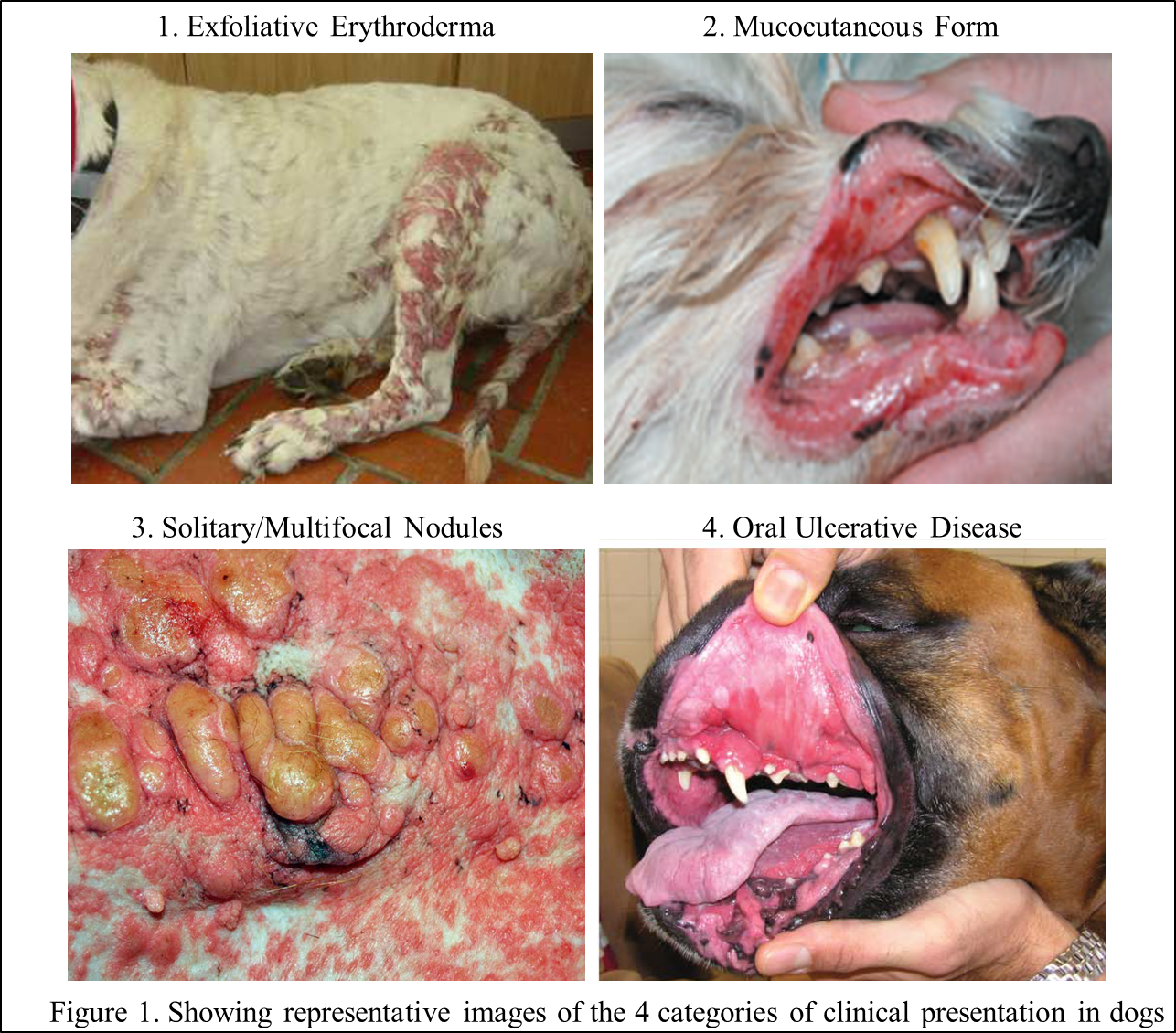
Clinical presentation of this condition is non-specific and can mimic many skin conditions. It affects skin and mucocutaneous junctions or oral mucosa. The clinical presentation is highly variable and hence it is classified into 4 different categories (Figure 1).
- Exfoliative erythroderma: Characterized by erythema over large areas of the body with scaling, depigmentation and alopecia; often these lesions will progress to plaques, patches and nodules. The distribution of lesions is generalized but most frequently involves head and trunk.
- Mucocutaneous Form: This form is common in dogs and often affects lips, nose and eyelids. The lesions are characterized by erythema, depigmentation, alopecia, irregular infiltration, erosion, and ulceration. Depigmentation is a common and clinically striking feature with marked bilateral symmetry.
- Solitary or multifocal cutaneous plaques or nodules: Characterized by solitary or multiple plaques or nodules that usually are erythematous and scaly or crusted. The larger lesions often erode and ulcerate. This stage can progress to neoplastic lymphadenopathy with eventual systemic spread.
- Ulcerative disease of oral mucosa: Oral mucosal involvement is not uncommon in dogs and can sometimes be limited to the oral mucosa; commonly affects gingiva, palate, or tongue that can progress to ulcerations.
Biopsy Sample Collection and Histopathology
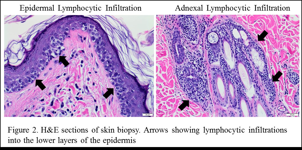
Epitheliotrophism cannot be identified by cytology - a definitive diagnosis is always based on histopathological examination. Hence, a full-thickness skin biopsy section is required for a definitive diagnosis. A characteristic infiltration of lymphocytes along the lower layers of the epidermis or mucosal epithelium and adnexal structures including the hair follicular walls and apocrine glands is considered diagnostic (Figure 2).
Prognosis:
The prognosis is often poor and is dependent on the clinical stage of presentation. The average age of survival is a few months to 2.5 years from the time of diagnosis. For more information on available treatment options please contact an oncologist at the Veterinary Health Center (VHC), College of Veterinary Medicine, Kansas State University. Please call KSVDL Client Care (866-512-5650) to setup an oncology consult with one of the oncologist at VHC.
Follow this link for “6 Tips for Biopsy Submissions”
Antigen Blocking and Heartworm Heat Treatment Antigen ELISA
Return to Index