
Maximizing the Diagnostic Potential of Skin Biopsies
By Dr. Gordon Andrews
Originally published in the September 2015 issue.
For dermatological cases, properly obtained and interpreted skin biopsies can be the cornerstone of establishing a definitive diagnosis, or a differential diagnosis list that can be refined by additional diagnostic testing. To be successful, this requires effort by both the submitter and pathologist. The following guidelines are suggested to ensure that your pathologist has appropriate biopsies to examine and clinical information to interpret the biopsies and put the histological lesions into context to establish a diagnosis.
STOP medication if possible: When feasible; withhold the patient from drugs, particularly steroids, prior to obtaining biopsies. Steroids are anti-inflammatory and can alter the histologic lesions and nature of the inflammatory cell infiltrate.
DO NOT prep the biopsy sites: The pathologist needs to see the crust, scale, or exudate on the skin surface. Crust, scale or exudate on the surface may be the only change in some skin diseases, or may be part of a combination of lesions in other diseases. Etiological agents such as mites, yeast or bacteria may be in the surface crust. Pustules and vesicles are fragile and easily ruptured by prepping the skin.
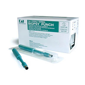
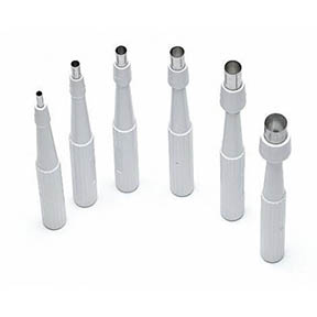
ALWAYS use sharp dissection to obtain biopsies: Skin punch biopsy instruments are preferred. They are inexpensive, easy to use and take uniform clean tissue samples. they come in sizes from 2-8 mm diameter.
Use the largest punch practical for the anatomic site. 8 mm is recommended for general use. Smaller biopsies may be needed for sensitive areas such as the nose, around the eyes, and feet. Small biopsy sites can be left open to heal. Larger sites can be closed with a single suture. If lesions have discrete borders, use a scalpel to take an elliptical biopsy with the long axis of the ellipse perpendicular to the edge of the biopsy. Never use laser or electrocautery to obtain skin biopsies. These instruments will burn tissue. Small skin biopsies can be completely burned and rendered useless for histopathology. Handle tissues gently. Crush artifact alters microscopic anatomy and destroys cellular detail. Label this biopsy as lesion border. Color differences that are grossly obvious can disappear when the tissue is fixed.
TAKE multiple biopsies: Multiple biopsies increase the probability of obtaining an accurate diagnosis. The diagnosis is seldom present in a single biopsy. Obtain biopsy samples from lesions that have differing gross appearances. Multiple biopsies may provide additional information about secondary problems like superficial pyoderma in addition to an immune mediated skin disease. In some cases (particularly diseases characterized by alopecia) a biopsy of clinically normal appearing skin can be helpful for comparison. Diagnostic microscopic lesions can be present in clinically normal appearing skin. Label this biopsy as normal appearing skin.
PUT the tissues in formalin as soon as possible: Desiccated tissue has altered staining characteristics and histologic appearance. Identify the anatomical location of each biopsy. Label each biopsy. Submitting the individual biopsies in tissue cassettes is ideal if you have them. These can be obtained from the diagnostic laboratory. Another method is to place the tissue in a gauze pouch and secure the pouch with string or suture.
CONSIDER performing additional diagnostic procedures: Perform a deep skin scrape to check for mites. Skin cytology can be performed by scraping or imprinting. Stain the slide and examine it yourself. Send an air-dried unstained slide to the lab for a clinical pathologist to examine. Would a bacterial or fungal culture be appropriate?
SHIPPING the sample: It is essential to ship this slide in a separate mailing container from the formalin fixed tissue. Formalin vapors from even sealed formalin containers fix the cells on the cytology slide and render the slide useless for cytologic examination. Ship the slide in a box to protect from breakage. DO NOT use cardboard slide mailers in an envelope. The slide will arrive broken. Obtain a fresh tissue biopsy for bacterial or fungal culture if you suspect infectious agents.
DOCUMENT the gross lesion(s): Today, digital cameras, computers, and web access are ubiquitous. Keep a digital camera in your exam room. Print the photos and send with the biopsy submission. Keep a set in the patient’s medical record. If your records are completely electronic, keep the digital files in the medical record. Alternatively, email digital images to us: receiving@vet.k-state.edu.
Indicate you are submitting biopsies to go with the photos. Identify the patient and owner and we will get the photos into the case file. Use your smart phone to take photos and email us with the same device.
FILL out the submission form completely: You should treat this case as a referral to a histopathologist. Fill out the submission form yourself. The more information your pathologist has, the more helpful they can be. Pertinent clinical information should include lesion distribution and gross characteristics (photos help greatly for this), duration and progression of lesions, presence of pruritus or pain, response or lack of response to therapy, specific drugs used, dose and duration of therapy if known, results of ancillary tests: (CBC, Chemistry panel, Endocrine testing, Cytology, Skin scrape), signs of systemic illness or other medical conditions (pancreatitis, liver disease, neoplasia, etc).
Dermatological conditions are among the most frequent presentations to companion animal practitioners. The skin is the largest organ of the body and is the easiest organ from which to obtain biopsy samples. Gross lesions are visible to the unaided eye without any specialized equipment and are easily documented by photography. Every veterinary practitioner has the ability to obtain high quality skin biopsies and provide a good clinical history so that the practitioner/pathologist team can establish an accurate as possible diagnosis.