
March 2019
Canine Ischemic Dermatopathy: Part II
Generalized Vaccine-Induced Ischemic Dermatopathy (GVIID)
(Continued from part I)
By Dr. Charan Ganta
|
|
|
Table 1 |
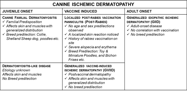
Canine ischemic dermatopathies include a group of ischemic skin diseases that result from cell poor vasculitis. These include juvenile, vaccine induced and adult onset types (Table 1). As a continued discussion of the ischemic dermatopathies in small animals here we present a case of generalized rabies vaccine-induced ischemic dermatopathy in a 7-year-old poodle after few months post rabies vaccination.
Condition and Clinical Manifestation
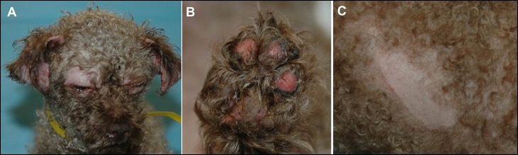
Generalized vaccine-induced ischemic dermatopathy is a post-vaccination skin disease characterized by clinical signs similar to familial dermatomyositis affecting skin and skeletal muscle. Skin lesions are characterized by alopecia, erythema and crusting predominantly affecting the areas of bony prominences like periocular skin, foot pads etc., where the areas are subjected to mechanical trauma. The areas of the skin lesions indicate underlying skeletal muscle atrophy. As can be seen in Figure 1, there is alopecia and erythema on the periorbital skin, foot pads and the site of vaccination.
Etiology and Pathogenesis
Generalized vaccine-induced ischemic dermatopathy cases will include a history of vaccination, most commonly with rabies vaccine. However, dermatopathy following polyvalent vaccine administration has also been reported. In addition, there is an increased risk for Miniature and Toy Poodle, Shih Tzu, Shetland Sheepdog, Lhasa Apso, Pomeranian, and Yorkshire Terrier breeds. Long-haired toy or miniature breeds seem to be at greater risk. It is believed that the vasculitis and myositis are secondary to deposition of rabies viral antigen complexes and complement (C5b-9/membrane attack complex) in the vessel walls and muscle.1
|
|
|
Figure 1. (A) Miniature poodle mix with vaccine-associated ischemic dermatopathy. Alopecia of the periocular skin and pinnal margins, and the ulceration/crusting of pinnal margins. (B) Ulcerative footpads of the patient in. (C) Rabies vaccination site showing alopecia and mild erythema. Image courtesy: Daniel O. Morris Vet Clin Small Anim 43 (2013) |
Disease Diagnosis and Sample Collection
The diagnosis of vaccine-based ischemia is based on the clinical presentation, vaccination history, and histopathology. Often the disease can be complicated by secondary conditions like pyoderma, demodicosis, dermatophytosis and Malassezia; therefore, multiple deep skin biopsy sections collected from different locations should be submitted.
Histopathological Features
The lesions are common in all conditions of canine ischemic dermatopathy, which is characterized as a cell-poor vasculitis (due to vascular compromise and cutaneous ischemia). The vascular changes include loss of endothelial cells, with hyalinization and leukocytoclasia. Altered staining of the collagen, fading atrophy of the follicles is also frequently noticed. In advanced cases, dermal-epidermal vacuolation and artefactual lifting of the epidermis could occur. Muscular lesions when noticed include mixed inflammation, regeneration, fibrosis, and atrophy.
Prognosis
This is a progressive disease and management of ischemic dermatopathies is often challenging and dependent on the stage and severity of the disease and the presence of secondary infections. I recommend calling the Kansas State University College of Veterinary Medicine Veterinary Health Center to setup a consultation with the clinical dermatologist at the Veterinary Health Center to discuss the management of this condition.
For any additional questions and references related to this newsletter please contact KSVDL Client Care at 866-512-5650 or clientcare@vet.k-state.edu
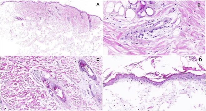 |
| Figure 2. H&E sections. Three-year-old Poodle with history of rabies vaccination. A. Diffuse and moderate to severe follicular atrophy with homogenization and pallor of dermal collagen. B. Cell poor vasculitis with vascular endothelial hyalinization. C. Marked follicular atrophy and pale staining dermal collagen. D. Dermal-epidermal junction showing cell-poor lymphocytic infiltration, with basal cell vacuolation. Images: Charan Ganta |
References:
1. Vaccine-induced ischemic dermatopathy in the dog. Carlo B. Vital, et al. Veterinary Dermatology 1999, 10, 131-142
Related articles:
- Canine Ischemic Dermatopathy
- An Injection Site Reaction: Post-Rabies Vaccination Panniculitis in a Toy Poodle