May 2020
Diagnosing Canine Lymphoma: When Cytology Alone Isn’t Enough
By Dr. Brandy Kastl
Cytologic evaluation of fine needle aspirate samples from enlarged peripheral lymph nodes is a relatively easy, minimally invasive, and inexpensive initial diagnostic tool when peripheral lymphoma is suspected. Readers are referred to an article for a review on cytologic evaluation of peripheral lymph nodes. However, cytologic evaluation has limitations that can hinder a definitive diagnosis of lymphoma (see Table 1). Even excellently prepared, highly cellular smears do not always yield a definitive diagnosis. Regardless of why, it still makes you want to shake your fist in the air and demand to know why you, your patient, and your client can’t catch a break. Let the frustration out. This article discusses the three most commonly employed diagnostic tests when a definitive cytologic diagnosis of canine lymphoma cannot be reached, 1) histopathologic evaluation, 2) immunophenotyping, and 3) PCR for antigen receptor rearrangement (PARR).
Table 1: A cytologic diagnosis of lymphoma can be hindered by:
- sampling of a lymph node that is not fully effaced by neoplastic lymphocytes
- disproportional sampling of a large reactive lymphoid nodule
- lymphomas comprised of predominately small lymphocytes (i.e. small cell lymphomas)
- prior administration of glucocorticoids reducing neoplastic lymphocyte numbers
- misinterpretation by the human evaluating the slides
- smear preparations lacking intact and/or well-spread cells
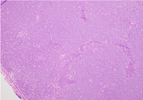
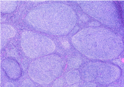
Despite the widespread use of cytology, histologic evaluation of nodal architecture remains the best method for differentiating reactive from neoplastic lymphoid populations. The exact histologic subtype, which can have prognostic implications, can also be determined. For instance, diffuse large B cell lymphoma (Figure 1) can be differentiated from follicular lymphoma (Figure 2). Both of these lymphomas are comprised of predominately larger B lymphocytes. However, canine follicular lymphoma has an indolent clinical course with a reported survival time sometimes longer than two years. In contrast, the reported average survival time for canine diffuse large B cell lymphoma is approximately 6 months. Histologic evaluation can also have several drawbacks, such as the invasiveness of surgical excision, some delay associated with tissue processing, and a higher relative cost. Furthermore, evaluation of standard hematoxylin and eosin stained sections alone cannot determine if lymphoma is of B or T cell origin. There are also instances when reactive and neoplastic lymphocyte populations cannot be differentiated. Immunophenotyping and PARR, respectively, address these limitations.
|
|
|
| Figure 1: Canine Diffuse Large B Cell Lymphoma; H&E stained, lymph node section, 10x objective | Figure 2: Follicular lymphoma in a human lymph node (histologically similar to canine follicular lymphoma). (image source) |
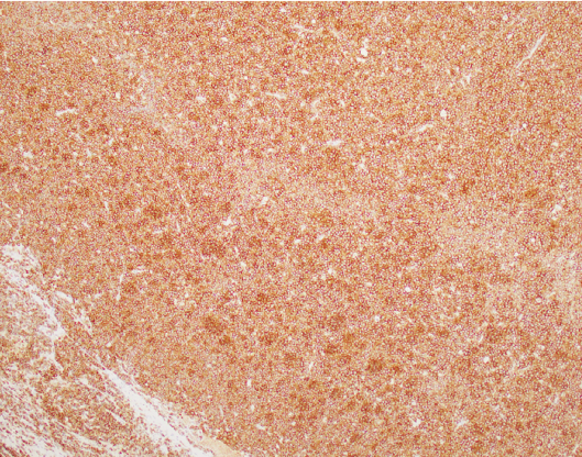
Immunophenotyping refers to the use of antibodies to detect protein expression in cells. Importantly, immunophenotyping is not a replacement for histologic and/or cytologic evaluation of peripheral lymph nodes. It also should not be used as a stand alone test to differentiate neoplastic and reactive lymphoid populations. Instead, immunophenotyping is an adjunct diagnostic tool used to determine the type of neoplastic lymhpocyte population present. In the context of canine lymphoma, it is most frequently used to determine if a lymphoma is of B or T cell origin. Canine lymphomas of T cell origin tend to have worse prognoses than those of B cell origin. Canine T-zone lymphoma is a notable exception to this rule as has an indolent clinical course with survival times often of two to four years reported in the literature. Immunophenotyping can be accomplished via immunohistochemical staining on histologic samples (Figure 3) or flow cytometry. Readers are referred to an article for further information on the use of flow cytometry for immunophenotyping.
|
|
| Figure 3: Canine Diffuse Large B Cell Lymphoma (same as Figure 1); Positive immunohistochemical staining for expression of CD20, B cell marker, indicated by the brown chromogen; lymph node section, 10x objective |
In contrast to immunophenotyping, PCR for antigen receptor rearrangement (PARR) is used to differentiate reactive and neoplastic lymphocyte populations. PARR is also an adjunct diagnostic test to cytologic and/or histologic evaluation. Importantly, it should not be used as a stand alone diagnostic in place of traditional assessment of cellular morphology and/or nodal architecture. Each lymphocyte has a unique genetic code for its’ antigen receptor – either a T-cell receptor for T cells or an immunoglobulin (antibody) receptor for B cells. Because neoplastic lymphoid populations are derived from clones of a single neoplastic lymphocyte, the presence of a single antigen receptor DNA sequence supports a diagnosis of lymphoma. However, this test does have some important limitations. First, a sufficient quantity of DNA representative of the entire lymphoid population must be available for testing in order to prevent false positive or false negative results. False positives have also been reported in cases of canine ehrlichiosis and tumors of macrophage/histiocytic origin. Uncommonly, lymphomas of T cell origin can have rearrangements of the immunoglobulin receptor gene sequence. Because of this, PARR should not be used to determine if a lymphoma is of B or T cell origin. Gene rearrangements in neoplastic cells do not always correlate with the proteins those cells express. Ultimately, it is the protein expression, ie the immunophenotype, that determines the cell of origin for clinical prognostication.
Cytologic evaluation of lymph node aspirates remains an easy, and often reliable, method for diagnosing peripheral canine lymphoma. Fortunately, several options are available when cytologic evaluation alone is insufficient for an accurate diagnosis. Histologic evaluation should be used to asses tissue architecture and determine the histologic subtype of lymphoma. Immunophenotyping, via immunohistochemical staining or flow cytometry, should be used to determine if lymphoma is of B or T cell origin. Finally, PARR can help distinguish reactive and neoplastic lymphocyte populations in conjunction with the cytologic and/or histologic findings. Together, all of these tests work to provide a complete picture for the prognosis for canine lymphoma patients.
Brandy Kastl, DVM is a diplomate of the American College of Veterinary Pathologists and a clinical pathologist in the Kansas State Veterinary Diagnostic Laboratory at Kansas State University.


