November 2019
Top 10 Urinalysis Tips
By Dr. Brandy Kastl
1. Love the gloves.
Leptospirosis is a bacterial disease transmitted to humans via the urine of infected animals. Affected individuals, both people and animals, can suffer from vomiting, acute renal failure, severe liver disease, and jaundice. In the last two years, the KSVDL identified 80 cases of leptospirosis in animals via our urine leptospira PCR. Because this organism is intermittently shed in urine, the true number of leptospirosis cases is likely higher. This disease is a serious zoonotic threat for veterinary professionals. Always wear gloves while handling animal urine. Wash your hands with warm water and soap when finished.
2. Collection method counts.
When possible, a minimum of 5 mL of urine should be collected. Ultrasound-guided cystocentesis is the best method for collection of a truly representative urine sample in small animals. If adequate time and technical skills are available in your practice, samples collected via sterile catheterization are also representative. Voided samples are often contaminated with environmental bacteria or protein from the lower urinary tract preventing an accurate diagnosis of urinary tract infections and/or renal protein loss. When a voided sample is necessary, collection from a clean, dry surface free of bleach-based cleaning solutions is recommended. Manual bladder expressions to collect voided samples are no longer recommended in patients with normal neurologic bladder tone and function. During manual expression, pressure on the bladder retropulses urine up into the kidneys which can spread bacterial infections from the lower urinary tract to the kidneys worsening the clinical disease and prognosis for our patients.
3. Remove all tube tops.
A new sterile red top (estimated cost/tube ~$0.10) or conical urine tube (estimated cost/tube ~$0.03) should be used for every sample. All urine samples should be handled under the assumption that a urine culture and sensitivity could be needed. Red tube stoppers and collection needles can serve as sources of environmental bacterial contamination. To prevent this, both rubber stoppers and needles should be removed before placing urine into the tube.
4. No time like the present.
For best results, urinalyses should be completed within 1 hour of collection. Otherwise, samples should be placed in a red top tube and refrigerated. With delayed processing or refrigeration, cells and bacteria continue to consumeglucose (sugar) in the urine falsely decreasing the urine glucose value on the dipstick. Bacteria will also produce ammonia making the urine more alkaline and increasing urine pH. This increased pH causes cells, like white blood cells, to degrade sometimes making them unrecognizable. All of these changes could result in an inaccurate interpretation and misdiagnosis.
5. Cold urine is unrelia ble urine.
Cold urine can falsely increase urine specific gravity measurements. Aritfactual amorphous crystals are often present in refrigerated urine samples, even those allowed to return to room temperature, and do not indicate disease. Every urinalysis should be performed on room-temperature urine. Refrigerated urine samples should sit at room temperature for 30 minutes before performing the urinalysis.
6. Reflect on your refractometer.
For the best results, a veterinary species-specific refractometer should be used. Several options which have been appropriately calibrated for both canine and feline urine samples are currently available. Refractometers should be calibrated with distilled (not tap) water which has a USG of 1.000 prior to every use. After each use, refractometers should be cleaned according to manufacturer instructions.
7. Dip the dipstick.
Some reagent pads on urine dipsticks are designed to absorb urine from along the sides of the pad rather than the top. Dipsticks should be completely immersed into the urine for ~1 second prior to sample centrifiguation. Use of conical urine tubes and thumb forceps (tweezers) may make dipping easier. Tap off excess urine. Read the color change for each pad following the appropriately designated reaction time of 30-60 seconds. Urine dipsticks were created for use in people. Results for these pads should be ignored due to frequent inaccuracy: leukocytes, nitrites, urobilinogen, and specific gravity.
8. Don’t skip the sediment.
Urine sedimentation is the only way to detect bacteria, white blood cells (leukocytes), urinary epithelial cells, and urinary crystals which are all important in the diagnosis of urinary tract disease. Prepare urine sediments by centrifuging ~5 mL of urine for 5 minutes. Pour or pipette off the excess urine leaving the pellet of cells and crystals at the bottom of the tube. Resuspend the pellet in the small volume of remaining urine.
|
|
|
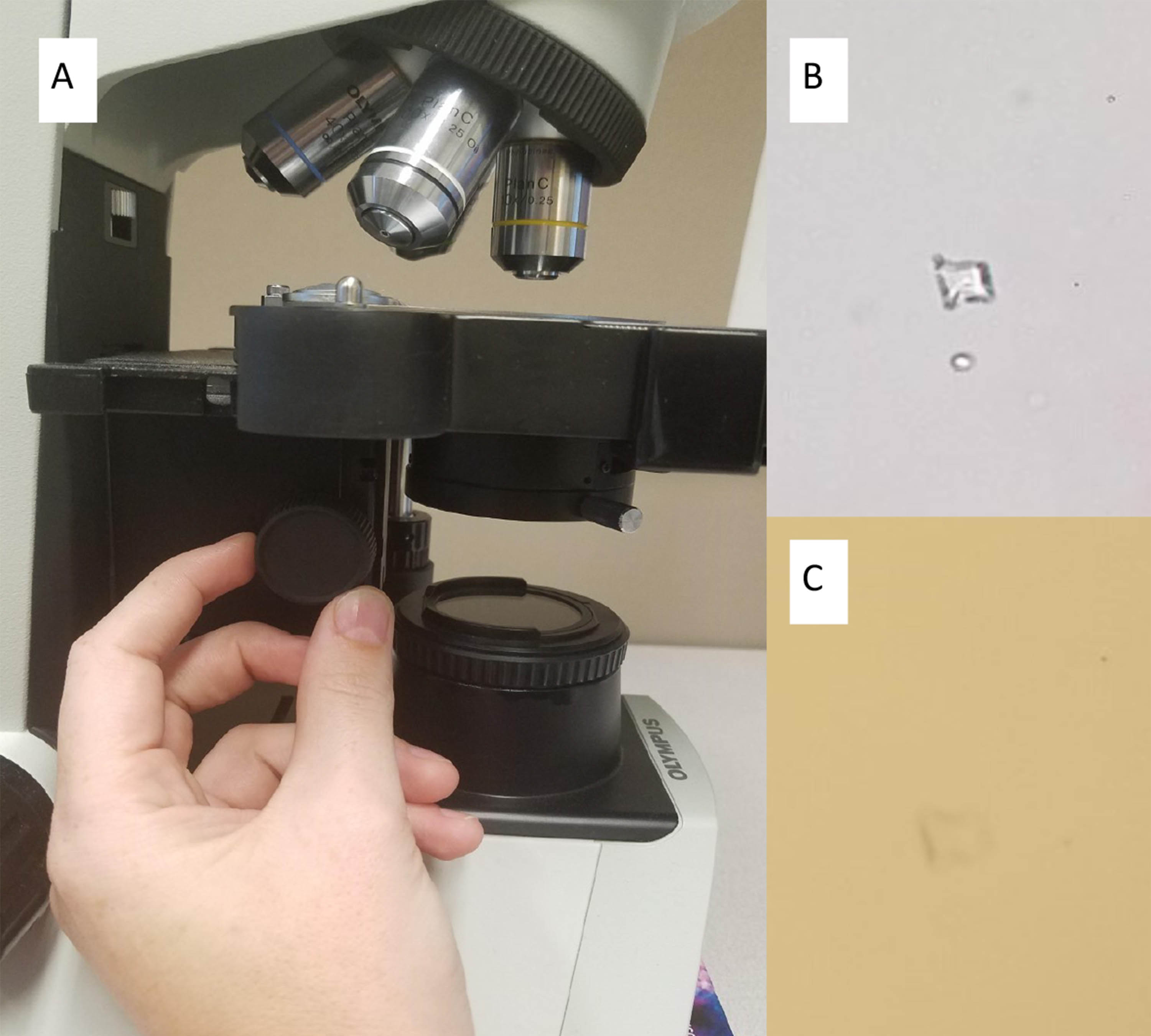
A) The condenser knob is typically on the left-hand side of the stage. Condenser is all the way up in this image. B) An easily visualized crystal when the condenser is down. C) The same crystal with condenser turned all the way up is fuzzy and difficult to see. |
9. Crystals pop when condensers drop.
An unstained wetmount preparation is best for visualization of urine crystals. Place a drop of the resuspended urine sediment pellet in the center of a clean, previously unused glass slide. Immediately cover with a clear coverslip. Drop the substage condenser to the lowest position which will allow crystals to refract the lower light against a darker background. Substage condensers are located underneath the microscope stage which the slide sits on.
10. Diff-Quik® makes the difference.
Diff-Quik® preparations are easy to make and better than SediStain® preparations for identifying bacteria and cells in the urine sediment. Pipette one drop of the resuspended urine sediment pellet onto one end of a new, clean glass slide. With a second new glass slide or coverslip, spread the droplet gently along the length of the slide similar to preparing a fine needle aspirate cytology smear. Allow the smear to air dry completely. Stain the smear according to manufacturer directions with Diff-Quik® or a similar in-house staining system. Be sure to return the condenser of the microscope up to just underneath the slide stage for easiest visualization of the cells.
Dr. Kastl (DVM, DACVP :Clinical Pathology) is a clinical pathologist in the Kansas State Veterinary Diagnostic Laboratory
