
September 2018
Canine Cutaneous Plasmacytoma: New Insights
By Drs. Rebecca Bacon and Chanran Ganta
What is a Plasmacytoma?
Canine extramedullary plasmacytoma is a benign, round-cell neoplasm that is derived from plasma cells of B cell lineage. A study consisting of 751 extramedullary plasmacytomas reported the most common location for this neoplasm was the skin (86%), and most frequently they were found on the head and limbs. Non-cutaneous sites include oral cavity (9%) and colon/rectal mucosa (4%). Most of these tumors are solitary, and <1% are associated with multiple myeloma (plasmacytoma arising in the bone marrow).
|
|
|
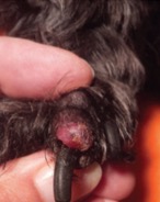
Figure 1. Toe, cutaneous lasmacytoma Gupta et. al. JAVMA, Vol 244, No. 2, January 15, 2014 |
Incidence and Known Biological Behavior:
Cutaneous plasmacytomas usually present clinically as alopecic, pink, raised, round, and well circumscribed growths of 1–2 cm in diameter (Figure 1). These are often reported in 5 to 11-year-old dogs with greatest breed predisposition for West Highland White Terriers, Golden Retrievers, Labrador Retrievers, Cocker Spaniels and Standard Poodles; however, plasmacytomas have also been reported in other breeds. Cutaneous plasmacytomas are benign and show no recurrence after complete surgical excision of the tumor; however, incompletely excised cutaneous neoplasms have been known to recur. Oral plasmacytomas are biologically more aggressive than cutaneous counterparts and can undergo local invasion into the underlying bone, however a complete surgical excision of the mass results in only rare recurrence or metastasis.
New Insights into Histopathological and Biological Behavior Correlation for Cutaneous Plasmacytoma:
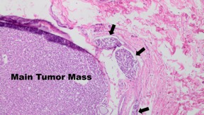
A recent study showed that approximately 16% of cutaneous plasmacytomas showed vascular invasion on histopathological examination (Figure 2). However, a follow-up study in these patients for up to 1 year showed no distant metastasis or local recurrence following complete surgical excision of the tumor. In addition, most of these neoplasms with intravascular invasion tend to be in the distal limbs (36%).
|
|
|
Figure 2. Plasmacytoma with vascular invasion shown in the peripheral thin vessels (arrows) |
In summary, cutaneous plasmacytomas are benign, often solitary, solid neoplasms offer good prognosis with complete surgical excision. Surprisingly, even with vascular invasion, recurrence or distant metastasis was not reported following complete surgical excision of the mass.
Please contact KSVDL if you have any questions or need references at clientcare@vet.k-state.edu
NEXT: Fall Discounts on Bovine Respiratory PCR Panels Return to Index