August 2020
Dermatophytosis – An Overview for Veterinary Nurses
By Christine Hackworth, RVT
|
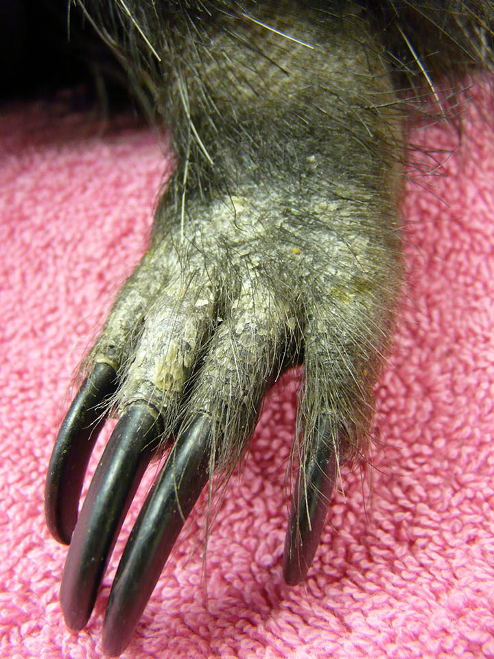
| Figure 1. Porcupine with ringworm |
Dermatophytosis, also known as ringworm, is a contagious fungal disease that can be zoonotic and transmissible from animal to animal and to humans. The most common organisms seen are Microsporium canis, Microsporium gypseum, and Trichophyton sp. Predisposing factors include very young and very old animals, stress, overcrowding, immunodeficiency, poor husbandry, malnutrition, concurrent underlying disease and pregnancy. 1,2 Although direct transmission between animals and people is considered uncommon, it cannot be completely ruled out. Animal handlers and owners need to know the importance of proper hygiene (hand washing) and the use of personal protective equipment (wearing disposable gloves).2,4 Proper disinfection of premises, using foot baths when going in and out of the enclosures and kennels, cleaning all surfaces with a disinfectant and removing all bedding and waste daily.2,4,5,7
Dermatophytosis should be suspected in animals showing lesions of alopecia, erythema, papules, scaling or crusting1,2,7 (Figure 1, courtesy of the Sunset Zoo). Remaining hairs may appear as stubble or broken.2,7 Common locations of dermatophyte lesions include the head, ears, and front limbs.7 The diagnosis of dermatophytosis is typically made by conventional techniques including trichography, fungal culture and histopathology.1-3,5,7 Use of the ultraviolet Wood’s lamp may reveal the presence of pathogenic dermatophytes (M. canis).1,2,5 A trichogram examines hairs under the microscope. The shafts and roots of the hair can be evaluated for spores or hyphae if fungus is present. Fungal culture is considered the most sensitive technique for the identification of dermatophytes.1 If fungal growth is observed on the culture media, the genus and species of the fungal organism can then be further identified microscopically.1,2,5 Positive fungal hyphae identification also enables the clinician to consider the likely source of the dermatophyte for subsequent control and protection of in-contact animals.1,2 Histopathology can help determine whether a fungal organism recovered in culture represents contamination, colonization or true infection.3 However, histopathology usually cannot identify the fungal genus and species, which are vital for specific diagnosis and treatment.3
Dermatophytosis is often a self-limiting disease with spontaneous resolution.1,2 However, medical therapy is typically warranted in order to successfully treat the animal and decrease the zoonotic potential of the disease.1,2,6,7 Treatment for dermatophytosis usually entails a multimodal approach and can include topical and/or systemic antifungal medications, environmental decontamination and careful hair clipping.2,4,5,7 Dermatophytosis may be treated with a variety of topical and systemic antifungal medications.1,2,5,7 For mild cases such as superficial, focal areas of infection, a cream or ointment can be applied to the affected areas. Prevention must be taken so that the animal does not lick off the medication.7 For animals that resist the application of topical medication or have a severe infection such as widespread, expanding lesions or deep infections accompanied by furunculosis, oral medications can be used. Treatment is typically recommended until at least two consecutive negative fungal cultures are obtained at one-to-two-week intervals.5
For a dermatophytosis case treated at the Veterinary Health Center in a porcupine, see this recent publication8 or the press release at http://www.k-state.edu/today/announcement.php?id=35631.
Christine graduated in 1999 from KSU with a bachelors of science degree in Life Science. In 2011 she graduated with an associate's degree from Penn Foster. She is currently an RVT in the Exotic and Dermatology sections of the Veterinary Health Center at the Kansas State University College of Veterinary Medicine.
References
- Bond R. Superficial veterinary mycoses. Clinics of Dermatology. 2010; 28:226-236.
- Dermatophytosis [Internet]. The Center for Food Security and Public Health; c2013 [cited 28 March 2016]. Available from: http://www.cfsph.iastate.edu/ Factsheets/pdfs/ dermatophytosis.pdf
- Guarner J, Brandt ME. Histopathologic diagnosis of fungal infections in the 21st century. Clinical Microbiology Reviews. 2011;24(2):247-280.
- Janssen DL. Guidelines for the management of zoonotic diseases. In: Miller RE, Fowler ME (eds.). Fowler's zoo and wild animal medicine current therapy. 8th ed. St. Louis (MO): Elsevier Saunders; 2015. p. 733-734.
- Moriello KA, DeBoer DJ. Treatment of dermatophytosis. In: Bonagura JD, Twedt DC (eds.) Kirk’s Current Veterinary Therapy XV. St. Louis, (MO): Elsevier; 2014. p. 449-451.
- Phair K, Larsen RS, Wack R. Dermatophytosis (Trichophytonmentagrophytes) in a Coquerel's Sifaka (Propithecuscoquereli). Journal of Zoo and Wildlife Medicine. 2001;42(4): 759–762.
- Pollock C. Fungal diseases of laboratory rodents. The Veterinary Clinics of North America: Exotic Animal Practice. 2003;6:401-413.
- Hackworth CE, Eshar D, Nau M, Bagladi-Swanson M, Andrews GA, Carpenter JW. Diagnosis and successful treatment of a potentially zoonotic dermatophytosis caused by Microsporum gypseum in a zoo-housed North American porcupine (Erethizon dorsatum). Journal of Zoo and Wildlife Medicine. 2017;48(2): 549-553.
Next: Blue-green Algae Season is Here!
Return to Index