May 2018
Equine Leukoencephalomalacia (ELEM)
By Drs. Giselle Cino, Mike Moore, Jonathon Sago and Jeffrey Laifer
Equine Leukoencephalomalacia is caused by the ingestion of moldy corn. The cerebral changes are caused by fumonisin compounds produced by Fusarium spp., a fungus that grows on corn grains. There are several different fumonisin compounds, however B1 is most commonly associated with ELEM.
Clinical History and Physical Examination Findings
An adult Appaloosa gelding was presented for gross necropsy. The night before, this horse had become ataxic, and was found dead the next morning. This horse was in a herd being fed grass hay and whole corn which was reported by the owner to be “slightly” moldy. At the request of their veterinarian, the owners had stopped feeding the moldy corn 3 weeks previous because another horse had presented with ataxia and blindness. Unfortunately, they began feeding the corn again, one week previous to the case reported here.
Gross Lesions
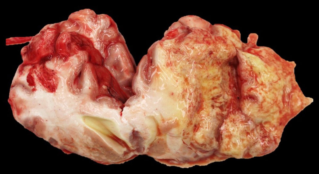
On necropsy, the only tissue affected was the brain. There was right-sided cerebral edema, yellow gelatinous malacia and liquefaction within the frontal, parietal, and temporal lobes. The areas of malacia spared the gray matter. Mild hemorrhage was present within the necrotic white matter. (Figure 1)
Microscopic Lesions
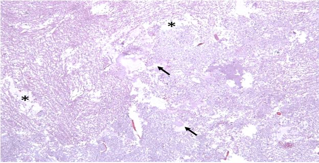
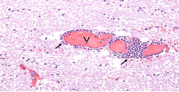
See Figures 2 and 3 (right column).
Laboratory Results
Rabies testing was negative.
Take Home Message
Equine Leukoencephalomalacia caused by ingestion of fumonisin can lead to severe neurological signs and death of multiple horses. Toxicity level and lesion severity depend on dose and length of time that horses are exposed to the affected feed; they are typically present 1-2 days after exposure. However, clinical signs may take weeks to develop occasionally. Clinical neurologic signs will depend on the severity of lesions and the anatomic locations most heavily affected. As there is no treatment for this condition, prevention measures are important and dependent on not feeding molding corn to horses.
References
-
Foreman, J.H., Constable, P.D., Waggoner, A. L., Levy, M., Eppley, R., Smith, G. W., Tumbleson, M.E. and Haschek, W.M. (2004). Neurologic abnormalities and Cerebrospinal Fluid Changes in Horses Administered Fumonisin B1 Intravenously. Journal of Veterinary Internal Medicine, 18: 223-230. Doi: 10.1111/j.1939-1676.2004.tb00165.x
-
Caloni, Francesco and Cortinovis, Cristina (2009), Effects of fusariotoxins in equine species. The veterinary Journal, 186, 157-161. Doi 10.1016/j.tvjl.209.09.020
-
McGavin, M.D. et al, Pathologic Basis of Veterinary Disease, (2012), pg. 843-844.
-
Mayhew, Ian G. Large animal neurology. 2nd ed., Wiley-Blackwell (2010), pg. 346-347
-
Blacutt, Alex A., et al. “Fusarium verticillioides: Advancements in Understanding the Toxicity, Virulence, and Niche Adaptations of a Model Mycotoxigenic Pathogen of Maize.” Phytopathology Review, 29 Sept. 2017, http://doi.org/10.1094/PHYTO-06-17-0203-RVW.
-
Giannitti F., Diab S.S., Pacin, A.M., Barrandeguy M., Larrere C., Ortega J. & Uzal F.A. 2011. Equine Leukoencephalomalacia (ELEM) due to fumonisins B1 and B2 in Argentina. Pesquisa Veterinaria Brasileira 31 (5): 407-412.
For more information contact KSVDL Client Care at 866-512-5650 or clientcare@vet.k-state.edu.


