November 2019
Feline Cutaneous Mast Cell Tumors: A newly proposed 2-tier grading system
By Dr. Charan Ganta
Incidence:

|
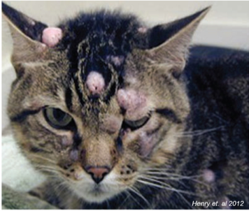
In cats, cutaneous mast cell tumors represent the second most common skin neoplasms and is composed of approximately 20% of all cutaneous neoplasms. The majority of these tend to occur on the head (~ 50%) followed by trunk (~35%) and limbs (~12%).
The existing literature characterizes feline cutaneous mast cell tumors as mostly benign, however approximately 20-25% of the cases show aggressive biological behavior with lymph node involvement and visceral metastasis. There is no grading system available in cats to distinguish these malignant cutaneous mast cell tumors from benign forms.
Proposed Grading Scheme:
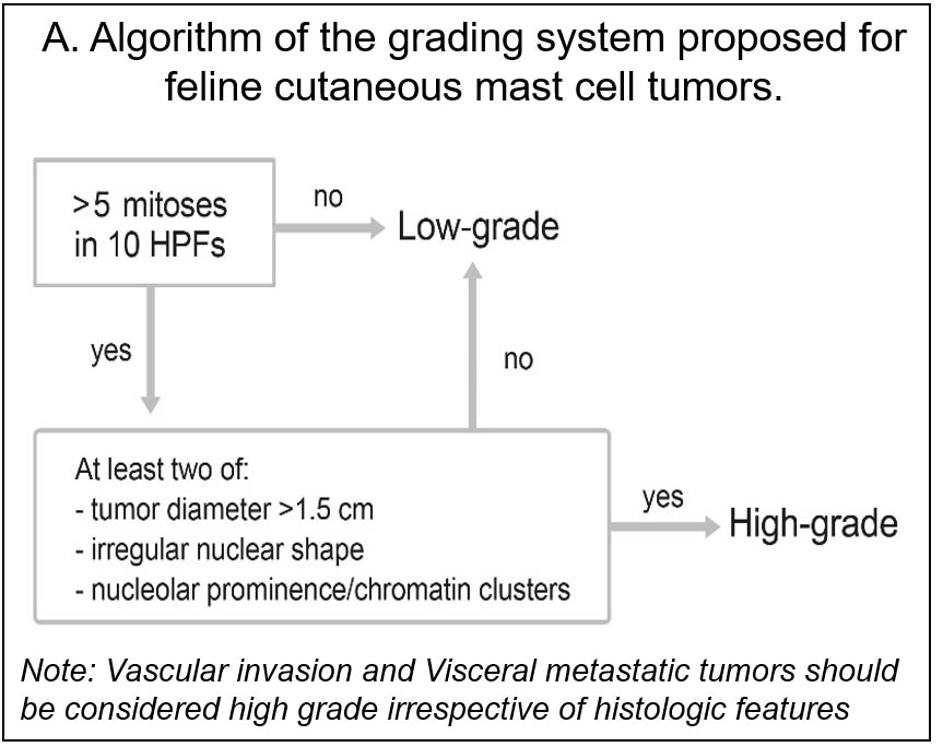
Mitotic count was considered to be prognostic factor for grading, however due to significant overlap in mitotic index between the benign and malignant forms, a new 2-teir grading system was proposed by Sabattini et. al., primarily based on mitotic index in combination with gross and histopathological features.
A high-grade tumor must have >5 mitotic figures in 10 HPFs and at least 2 of the following features: 1. Tumor diameter >1.5 cm; 2. Irregular nuclear shape; 3. Prominent nucleolus/chromatin clusters (Figure A).
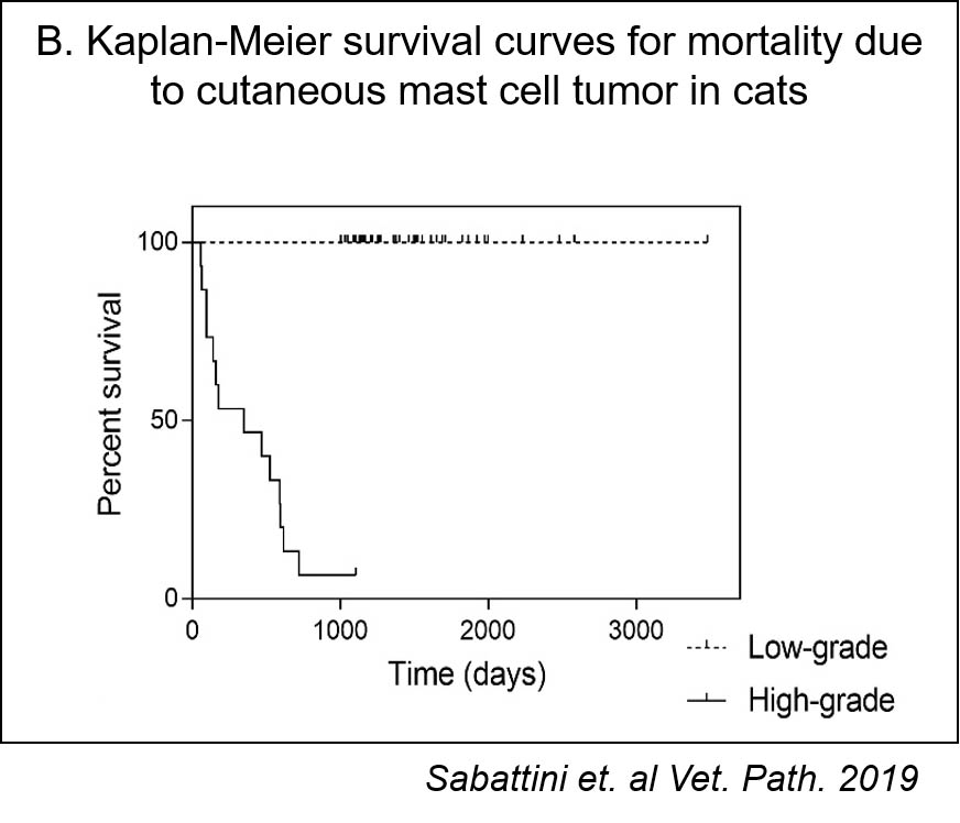
Based on the new grading scheme low grade mast cell tumors had prolonged survival (median not reached) and high-grade mast cell tumors had significantly decreased survival period (median, 349 days; 95% CI, 0–739 days), suggestive of significant correlation between the histologic grading and survival (Figure B).

|

|
Dr. Charan Ganta (BVSc, PhD, DACVP) is an anatomic pathologist at Kansas State Veterinary Diagnostic Laboratory.